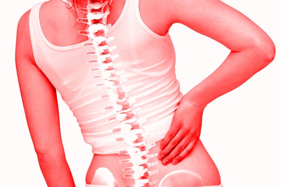
The spine (or spine) consists of vertebrae in the middle of which is a channel: the spinal canal. It contains the spinal cord and the nerves. At the lumbar level, the spinal cord stops and the canal contains only the nerves of the “ponytail” which innervate the lower limbs and the perineum.
This channel is usually wide enough to easily contain all nerve roots. Depending on the constitution of the patient or because of natural wear phenomena (osteoarthritis), a narrowing of the spinal canal (lumbar ductal stenosis or narrowed lumbar canal) may occur.
This narrowing or “stenosis” of the spinal canal will cause compression of the nerve elements contained in the spinal canal
Surgery alone widens the diameter of the spinal canal.
An intervention is considered when the patient has had a complete medical treatment, when he “has tired of having pain” or in case of emergency:
Motor deficit (paralyzing sciatica);
Intolerable pain not relieved by opioids (hyperalgic sciatica);
Syndrome of the ponytail (perineal disorders, sphincter dysfunctions especially urinary).

QU’EST-CE QUE L’ARTHROSCOPIE DE L’ÉPAULE
A la clinique Avicenne, la plupart des affections de l’épaule sont traitées par voie arthroscopique. Via arthroscopie ou « endoscopie », on introduit une caméra dans le cartilage. Le chirurgien traite alors la source de votre problème à l’épaule en pénétrant dans l’articulation grâce à une technique microchirurgicale.
Cette technique permet de parvenir à une solution définitive avec un minimum d’effets secondaires. Pour ce faire, on procède à quelques petites incisions de 5 à 10 mm autour de l’articulation de l’épaule. Ces dernières ne donnent que rarement voire jamais lieu à la formation d’une cicatrice.
QUELLES AFFECTIONS PEUVENT ÊTRE TRAITÉES GRÂCE À L’ARTHROSCOPIE DE L’ÉPAULE?
La plupart des affections de l’épaule peuvent être traitées grâce à une intervention arthroscopique. Ce n’est qu’en cas de nécessité de placement d’une prothèse due à une arthrose avancée que cette intervention doit être réalisée en technique ouverte. La plupart des fractures osseuses doivent également être traitées en chirurgie ouverte. À de rares exceptions près, TOUTES les opérations de l’épaule sont réalisées par voie arthroscopique.
Les opérations de l’instabilité de l’épaule (suite à des luxations de l’épaule) ne sont plus elles aussi réalisées que par voie arthroscopique depuis 1998. Le taux de réussite de ces interventions est au moins aussi élevé que les opérations en chirurgie ouverte réalisées par le passé.
QU’EST-CE QUE L’ARTHROSCOPIE?
Arthroscopie signifie littéralement « observer dans l’articulation ». Nous parlons ici d’une intervention chirurgicale où l’on observe et ou l’on travaille à l’intérieur d’une articulation à l’aide d’une mini-caméra.
C’est pour cette raison que les interventions arthroscopiques sont également parfois appelées endoscopies.
On ne fait pas qu’observer lors d’une arthroscopie ou d’une endoscopie. Les opérations chirurgicales sont réalisées au sein de l’articulation directement via la caméra. Il s’agit donc d’une notion très large.
Le point commun de l’ensemble de ces opérations est qu’on n’ouvre plus l’articulation.
L’arthroscopie est une opération spécifique des articulations. Elle appartient au groupe des « opérations endoscopiques.
Il existe également des opérations endoscopiques orthopédiques (donc en dehors de l’articulation)
QUELLES ARTICULATIONS PEUVENT ÊTRE TRAITÉES GRÂCE À L’ARTHROSCOPIE?
En principe, toutes les articulations peuvent être traitées par arthroscopie. De fait, la plupart des opérations des articulations du genou et des épaules sont depuis longtemps exclusivement exécutées par voie arthroscopique.
Cependant, ces dernières années l’arthroscopie du genou, du poignet, de la hanche et de la cheville sont également devenues plus courantes.
CONSÉQUENCES DE L’ARTHROSCOPIE
Il y a peu d’inconvénients ou de conséquences à une arthroscopie. Étant donné que l’articulation reste fermée, les tissus environnants ne subissent que peu voire aucun dommage, et le risque d’infection de l’articulation est extrêmement mince.
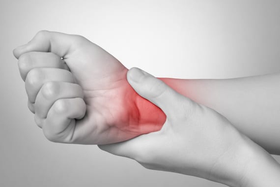
Carpal tunnel syndrome is the clinical translation of compression of the median nerve at the wrist. This nerve innervates the hand on the sensory plane (thumb, index, middle and half of the ring finger) and motor (muscles of the base of the thumb).
The nerve and flexor tendons pass through a bony canal (the carpal tunnel) which is covered with a thick ligament (the anterior ring ligament). Any increase in pressure in the carpal tunnel causes nerve pain.
Treatment
Symptoms may decrease with medical treatment. In case of associated pathology (diabetes, hypothyroidism, …), the treatment of the disease usually relieves carpal tunnel syndrome.
The rest of the hand concerned, a correct working position, or even wearing a brace of rest can help reduce the compression of the median nerve.
Intra-ductal infiltration can also relieve symptoms.
If signs persist or worsen, surgical treatment should be used. The purpose of the surgery is to cut the ligament covering the nerve, to reduce the pressure. The consequences are usually simple, the patient can resume a light activity in the week following the intervention. The force recovers in 6 to 12 weeks. Healing of the incision is complete in 15 to 21 days.
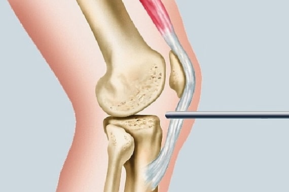
The cervical spine (or cervical spine) consists of 7 vertebrae stacked and separated by disks, except between the 1st and 2nd cervical vertebra (C1 and C2).
In the middle of the cervical spine is the spinal canal containing the spinal cord.
The normal intervertebral disk is a flat, cylindrical structure uniting the two vertebrae and acting as a shock absorber.
The disc deterioration is characterized by tears or fissures of the fibrous ring. The causes are dehydration due to aging, microtrauma due to mobility constraints and sometimes also traumas such as cervical sprains.
WHAT ARE THE OBJECTIVES OF SURGERY?
The purpose of the procedure is to remove the herniated disc to release the nerve root (or even the spinal cord) and to remove the pain in the upper limb
This goal is achieved in about 90% of cases. On the other hand, it can persist neck pain
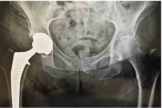
The implementation of a total hip replacement is suggested by the doctor when the disability has become severe and when joint damage is very advanced.
The patient needs to know what the intervention is and how it is exposed in the aftermath.
After being well informed, he decides. “Total” means that the prosthesis concerns the 2 parts of the hip joint: the part of the pelvis (the acetabulum) and the part of the femur (the head).
The models of total prosthesis are not lacking. They vary according to the shape, the material and the method of fixation of the prosthesis.
Pain that has not been sufficiently relieved by antalgic medications and the resulting handicap (walking problems, reduced activity) are good reasons to consider the introduction of a total hip prosthesis.
If, in addition, the osteoarthritis lesions are very advanced on the most recent radiographs, it is an additional reason to think about it.
The lifespan of a prosthesis varies from 15 to 25 years depending on the age, weight and activity of the patient.
Sorry, this entry is only available in French and Arabic.